Overview
The foot only contains one naturally occurring bursal sac between the Achilles tendon and the heel bone, which protects the Achilles from the pressure against the heel bone during ambulation. However, shoes may put pressure on this bursa. The bursa might also incur trauma from walking on hard ground. And, though they are not naturally occurring, bursa sacs can also form, and become inflamed, in other parts of the foot, including the bottom of the heel, and the metatarsal plate, the outside of the foot below the fifth toe, and so on.
Causes
Overuse of the ankle joint may cause irritation of the bursa such as excessive walking, running or jumping. Poor biomechanics and foot function may ultimately lead to heel bursitis due to pulling on the back of the heel by the Achilles tendon.
A sudden increase in physical activity without adequate rest may result in heel bursitis. Excessive standing and walking bare foot on hard surfaces.
Symptoms
Retrocalcaneal bursitis is very similar to Achilles bursitis as the bursae are very close in proximity and symptoms are almost identical however retrocalcaneal bursitis is a lot more common. The symptoms of bursitis vary depending on whether the bursitis is the result of injury or an underlying health condition or from infection. From normal overuse and injury the pain is normally a constant dull ache or burning pain at the back of the heel that is aggravated by any touch, pressure like tight shoes or movement of the joint. There will normally be notable swelling around the back of the heel. In other cases where the bursa lies deep under the skin in the hip or shoulder, swelling might not be visible. Movement of the ankle and foot will be stiff, especially in the mornings and after any activity involving the elbow. All of these symptoms are experienced with septic bursitis with the addition of a high temperature of 38?C or over and feverish chills. The skin around the affected joint will also appear to be red and will feel incredibly warm to the touch. In cases of septic bursitis it is important that you seek medical attention. With injury induced bursitis if symptoms are still persisting after 2 weeks then report to your GP.
Diagnosis
The diagnosis is based on the symptoms and an examination. For anterior Achilles tendon bursitis, doctors use x-rays to rule out a fracture of the heel bone or damage to the heel bone caused by rheumatoid arthritis or other inflammatory arthritis.
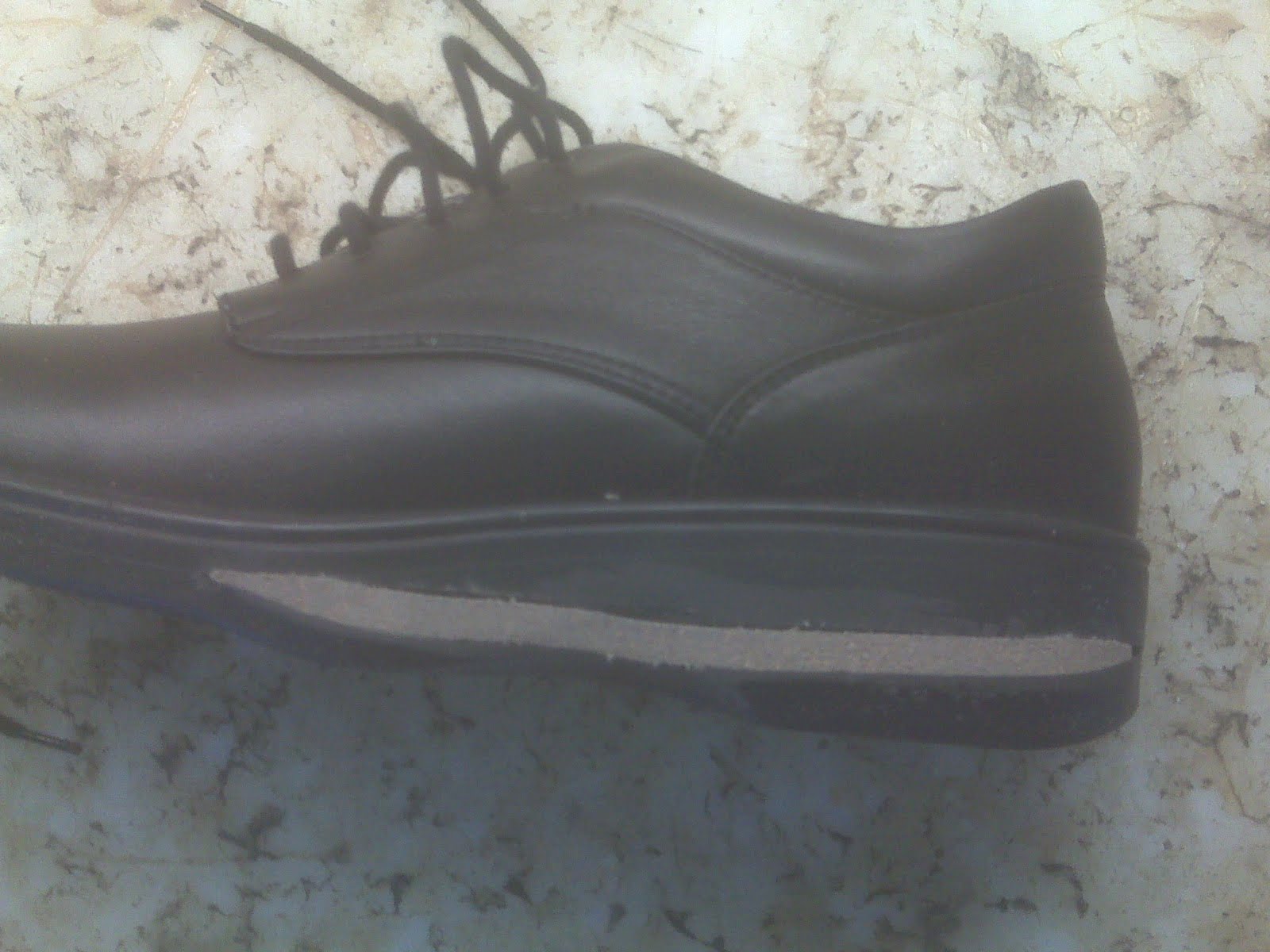
Non Surgical Treatment
Surgery should always be the last option. We believe that biologic treatments that preserve normal anatomy are very helpful, particularly for runner, athletes, and active professionals with buy schedules. All non-surgical approaches attempt to calm down the inflammation of the bursa and Achilles tendon. They do not address the bony bump, but they can substantially reduce and shrink the inflamed soft tissue. Some non-surgical treatments include Oral Anti-inflammatory Medications. NSAID's (non-steroidal anti-inflammatory medications) such as Motrin, Aleve, and Steroids (like prednisone) may help control the pain and stop the inflammation. Topical Anti-inflammatory Medications. NSAID's in cream or lotion form may be applied directly to the inflamed area. With these, there is no concern for stomach upset or other problems associated with oral medication. Ice. Ice can applied be applied right to the red, inflamed area and help calm it down. Try applying a podiatrist-approved ice pack to the affected area for 20 minutes of each hour. Just make sure you don't put ice directly against the skin. Exercises. Stretching exercises may relieve some of the tension in the Achilles tendon that started the problem. If you have Equinus Deformity (or a tight heel cord) this is critical to prevent it from coming back again. Heel lifts. Heel lifts placed inside the shoe can decrease the pressure on the Achilles tendon. Remember, pressure and friction cause the bump to become inflamed. Heel pads. Placing gel padding to cushion the Achilles tendon (at the back of the heel) can also help reduce irritation from shoes. Shoe modification. Wearing open-backed shoes, or shoes that have soft backs. This will also help stop the irritation. Physical therapy. Physical therapy, such as ultrasound, massage and stretching can all reduce the inflammation without surgery. Orthotic devices. Custom arch supports known as foot orthotics control abnormal motion in the foot that can allow the heel to tilt over and rub against the heel counter. Orthotics can decrease symptoms and help prevent it from happening again. Immobilization. In some cases, a walking cast boot or plaster/fiberglass cast is necessary to take pressure off the bursa and tendon, while allowing the area to calm down. ESWT. Extra-corporeal Shock Wave Therapy uses high energy sound waves to break up diseased tissue in the bursa and Achilles tendon and stimulate your own bodies healing processes to repair the diseased area. It may be done in the office or in a an outpatient surgery center. There is no incision and no stitches with ESWT. PRP. Platelet Rich Plasma (PRP) is a therapeutic injection. A small sample of blood is drawn from the patient and the healing factors found in the platelets are concentrated in a centrifuge. By injecting the concentrated solution right into the damaged Achilles tendon, a powerful healing can be stimulated. This can be done in the office. No hospital or surgery required.
Surgical Treatment
Surgery is rarely done strictly for treatment of a bursitis. If any underlying cause is the reason, this may be addressed surgically. During surgery for other conditions, a bursa may be seen and removed surgically.
Prevention
You can help to prevent heel pain and bursitis by maintaining a healthy weight, by warming up before participating in sports and by wearing shoes that support the arch of the foot and cushion the heel. If you are prone to plantar fasciitis, exercises that stretch the Achilles tendon (heel cord) and plantar fascia may help to prevent the area from being injured again. You also can massage the soles of your feet with ice after stressful athletic activities. Sometimes, the only interventions needed are a brief period of rest and new walking or running shoes.
The foot only contains one naturally occurring bursal sac between the Achilles tendon and the heel bone, which protects the Achilles from the pressure against the heel bone during ambulation. However, shoes may put pressure on this bursa. The bursa might also incur trauma from walking on hard ground. And, though they are not naturally occurring, bursa sacs can also form, and become inflamed, in other parts of the foot, including the bottom of the heel, and the metatarsal plate, the outside of the foot below the fifth toe, and so on.
Causes
Overuse of the ankle joint may cause irritation of the bursa such as excessive walking, running or jumping. Poor biomechanics and foot function may ultimately lead to heel bursitis due to pulling on the back of the heel by the Achilles tendon.
A sudden increase in physical activity without adequate rest may result in heel bursitis. Excessive standing and walking bare foot on hard surfaces.
Symptoms
Retrocalcaneal bursitis is very similar to Achilles bursitis as the bursae are very close in proximity and symptoms are almost identical however retrocalcaneal bursitis is a lot more common. The symptoms of bursitis vary depending on whether the bursitis is the result of injury or an underlying health condition or from infection. From normal overuse and injury the pain is normally a constant dull ache or burning pain at the back of the heel that is aggravated by any touch, pressure like tight shoes or movement of the joint. There will normally be notable swelling around the back of the heel. In other cases where the bursa lies deep under the skin in the hip or shoulder, swelling might not be visible. Movement of the ankle and foot will be stiff, especially in the mornings and after any activity involving the elbow. All of these symptoms are experienced with septic bursitis with the addition of a high temperature of 38?C or over and feverish chills. The skin around the affected joint will also appear to be red and will feel incredibly warm to the touch. In cases of septic bursitis it is important that you seek medical attention. With injury induced bursitis if symptoms are still persisting after 2 weeks then report to your GP.
Diagnosis
The diagnosis is based on the symptoms and an examination. For anterior Achilles tendon bursitis, doctors use x-rays to rule out a fracture of the heel bone or damage to the heel bone caused by rheumatoid arthritis or other inflammatory arthritis.
Non Surgical Treatment
Surgery should always be the last option. We believe that biologic treatments that preserve normal anatomy are very helpful, particularly for runner, athletes, and active professionals with buy schedules. All non-surgical approaches attempt to calm down the inflammation of the bursa and Achilles tendon. They do not address the bony bump, but they can substantially reduce and shrink the inflamed soft tissue. Some non-surgical treatments include Oral Anti-inflammatory Medications. NSAID's (non-steroidal anti-inflammatory medications) such as Motrin, Aleve, and Steroids (like prednisone) may help control the pain and stop the inflammation. Topical Anti-inflammatory Medications. NSAID's in cream or lotion form may be applied directly to the inflamed area. With these, there is no concern for stomach upset or other problems associated with oral medication. Ice. Ice can applied be applied right to the red, inflamed area and help calm it down. Try applying a podiatrist-approved ice pack to the affected area for 20 minutes of each hour. Just make sure you don't put ice directly against the skin. Exercises. Stretching exercises may relieve some of the tension in the Achilles tendon that started the problem. If you have Equinus Deformity (or a tight heel cord) this is critical to prevent it from coming back again. Heel lifts. Heel lifts placed inside the shoe can decrease the pressure on the Achilles tendon. Remember, pressure and friction cause the bump to become inflamed. Heel pads. Placing gel padding to cushion the Achilles tendon (at the back of the heel) can also help reduce irritation from shoes. Shoe modification. Wearing open-backed shoes, or shoes that have soft backs. This will also help stop the irritation. Physical therapy. Physical therapy, such as ultrasound, massage and stretching can all reduce the inflammation without surgery. Orthotic devices. Custom arch supports known as foot orthotics control abnormal motion in the foot that can allow the heel to tilt over and rub against the heel counter. Orthotics can decrease symptoms and help prevent it from happening again. Immobilization. In some cases, a walking cast boot or plaster/fiberglass cast is necessary to take pressure off the bursa and tendon, while allowing the area to calm down. ESWT. Extra-corporeal Shock Wave Therapy uses high energy sound waves to break up diseased tissue in the bursa and Achilles tendon and stimulate your own bodies healing processes to repair the diseased area. It may be done in the office or in a an outpatient surgery center. There is no incision and no stitches with ESWT. PRP. Platelet Rich Plasma (PRP) is a therapeutic injection. A small sample of blood is drawn from the patient and the healing factors found in the platelets are concentrated in a centrifuge. By injecting the concentrated solution right into the damaged Achilles tendon, a powerful healing can be stimulated. This can be done in the office. No hospital or surgery required.
Surgical Treatment
Surgery is rarely done strictly for treatment of a bursitis. If any underlying cause is the reason, this may be addressed surgically. During surgery for other conditions, a bursa may be seen and removed surgically.
Prevention
You can help to prevent heel pain and bursitis by maintaining a healthy weight, by warming up before participating in sports and by wearing shoes that support the arch of the foot and cushion the heel. If you are prone to plantar fasciitis, exercises that stretch the Achilles tendon (heel cord) and plantar fascia may help to prevent the area from being injured again. You also can massage the soles of your feet with ice after stressful athletic activities. Sometimes, the only interventions needed are a brief period of rest and new walking or running shoes.
 Overview
 Symptoms
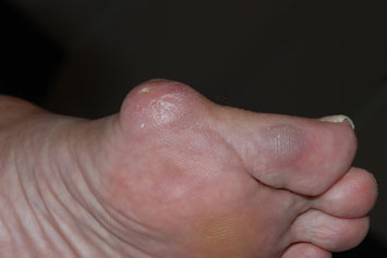 A bunion is an enlargement of bone or tissue around the joint at the base of the big toe. The big toe may turn toward the second toe. The tissues around the joint may be swollen and tender. A bony bump at the base of the little toe is called a bunionette or tailor's bunion. The little toe also bends inward, and the joint swells or enlarges.

 Causes Adult flatfoot typically occurs very gradually. If often develops in an obese person who already has somewhat flat feet. As the person ages, the tendons and ligaments that support the foot begin to lose their strength and elasticity. Symptoms Posterior tibial tendon insufficiency is divided into stages by most foot and ankle specialists. In stage I, there is pain along the posterior tibial tendon without deformity or collapse of the arch. The patient has the somewhat flat or normal-appearing foot they have always had. In stage II, deformity from the condition has started to occur, resulting in some collapse of the arch, which may or may not be noticeable. The patient may feel it as a weakness in the arch. Many patients initially present in stage II, as the ligament failure can occur at the same time as the tendon failure and therefore deformity can already be occurring as the tendon is becoming symptomatic. In stage III, the deformity has progressed to the extent where the foot becomes fixed (rigid) in its deformed position. Finally, in stage IV, deformity occurs at the ankle in addition to the deformity in the foot. Diagnosis Perform a structural assessment of the foot and ankle. Check the ankle for alignment and position. When it comes to patients with severe PTTD, the deltoid has failed, causing an instability of the ankle and possible valgus of the ankle. This is a rare and difficult problem to address. However, if one misses it, it can lead to dire consequences and potential surgical failure. Check the heel alignment and position of the heel both loaded and during varus/valgus stress. Compare range of motion of the heel to the normal contralateral limb. Check alignment of the midtarsal joint for collapse and lateral deviation. Noting the level of lateral deviation in comparison to the contralateral limb is critical for surgical planning. Check midfoot alignment of the naviculocuneiform joints and metatarsocuneiform joints both for sag and hypermobility. Non surgical Treatment The adult acquired flatfoot is best treated early. Accurate assessment by your doctor will determine which treatment is suitable for you. Reduce your level of activity and follow the RICE regime. R - rest as often as you are able. Refrain from activity that will worsen your condition, such as sports and walking. I - ice, apply to the affected area, ensure you protect the area from frostbite by applying a towel over the foot before using the ice pack. C - compression, a Tubigrip or elasticated support bandage may be applied to relieve symptoms and ease pain and discomfort. E - elevate the affected foot to reduce painful swelling. You will be prescribed pain relief in the form of non-steroidal antiinflammatory medications (if you do not suffer with allergies or are asthmatic). Immobilisation of your affected foot - this will involve you having a below the knee cast for four to eight weeks. In certain circumstances it is possible for you to have a removable boot instead of a cast. A member of the foot and ankle team will advise as to whether this option is suitable for you. Footwear is important - it is advisable to wear flat sturdy lace-up shoes, for example, trainers or boots. This will not only support your foot, but will also accommodate orthoses (shoe inserts).
  Surgical Treatment For those patients with PTTD that have severe deformity or have not improved with conservative treatments, surgery may be necessary to return them to daily activity. Surgery for PTTD may include repair of the diseased tendon and possible tendon transfer to a nearby healthy tendon, surgery on the surrounding bones or joints to prevent biomechanical abnormalities that may be a contributing factor or both.

 A rupture of the Achilles tendon means that there has been either a complete, or partial, tear of the tendon which connects the calf muscles to the heel bone. Usually this occurs just above insertion on the heel bone, although it can happen anywhere along the course of the tendon. Achilles tendon rupture occurs in people that engage in strenuous activity, who are usually sedentary and have weakened tendons, or in people who have had previous chronic injury to their Achilles tendons. Previous injury to the tendon can be caused by overuse, improper stretching habits, worn-out or improperly fitting shoes, or poor biomechanics (flat-feet). The risk of tendon rupture is also increased with the use of quinolone antibiotics (e.g. ciprofloxacin, Levaquin). Causes Achilles tendon ruptures are most likely to occur in sports requiring sudden stretching, such as sprinting and racquet sports. Achilles tendon ruptures can happen to anyone, but are most likely to occur to middle age athletes who have not been training or who have been doing relatively little training. Common sporting activities related to Achilles tendon rupture include, badminton, tennis, squash. Less common sporting activities that can lead to Achilles tendon rupture include: TKD, soccer etc. Occasionally the sufferer may have a history of having had pain in the Achilles tendon in the past and was treated with steroid injection to around the tendon by a doctor. This can lead to weakening of the tendon predisposing it to complete rupture. Certain antibiotics taken by mouth or by intravenous route can weaken the Achilles tendon predisposing it to rupture. An example would be the quinolone group of antibiotics. An common example is Ciprofloxacin (or Ciprobay). Symptoms A classic sign of an Achilles tendon rupture is the feeling of being hit in the Achilles are. There is often a "pop" sound. There may be little pain, but the person can not lift up onto his toes while weight bearing. Diagnosis In diagnosing an Achilles tendon rupture, the foot and ankle surgeon will ask questions about how and when the injury occurred and whether the patient has previously injured the tendon or experienced similar symptoms. The surgeon will examine the foot and ankle, feeling for a defect in the tendon that suggests a tear. Range of motion and muscle strength will be evaluated and compared to the uninjured foot and ankle. If the Achilles tendon is ruptured, the patient will have less strength in pushing down (as on a gas pedal) and will have difficulty rising on the toes. The diagnosis of an Achilles tendon rupture is typically straightforward and can be made through this type of examination. In some cases, however, the surgeon may order an MRI or other advanced imaging tests. Non Surgical Treatment The most widely used method of non-surgical treatment involves the use of serial casting with gradual progression from plantar flexion to neutral or using a solid removable boot with heel inserts to bring the ends of the tendon closer together. The advantage of a solid removable boot is that it allows the patient to begin early motion and is removable. Wide variability exists among surgeons in regards to the period of absolute immobilization, initiating range of motion exercises, and progression of weight bearing status.
 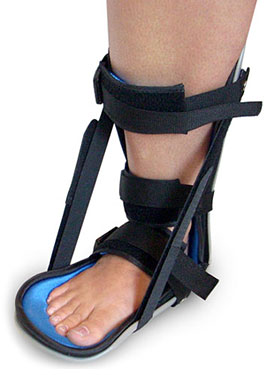 Surgical Treatment Surgery offers important potential benefits. Besides decreasing the likelihood of re-rupturing the Achilles tendon, surgery often increases the patient?s push-off strength and improves muscle function and movement of the ankle. Various surgical techniques are available to repair the rupture. The surgeon will select the procedure best suited to the patient. Following surgery, the foot and ankle are initially immobilized in a cast or walking boot. The surgeon will determine when the patient can begin weight bearing. Complications such as incision-healing difficulties re-rupture of the tendon, or nerve pain can arise after surgery. Prevention To help reduce your chance of getting Achilles tendon rupture, take the following steps. Do warm-up exercises before an activity and cool down exercises after an activity. Wear proper footwear. Maintain a healthy weight. Rest if you feel pain during an activity. Change your routine. Switch between high-impact activities and low-impact activities. Strengthen your calf muscle with exercises.
 The Achilles tendon is the confluence of the independent tendons of the gastrocnemius and soleus, which fuse to become the Achilles tendon approximately 5 to 6 cm proximal to its insertion on the posterior surface of the calcaneus. The gastrocnemius and soleus muscles, via the Achilles tendon, function as the chief plantarflexors of the ankle joint. This musculotendinous unit provides the primary propulsive force for walking, running, and jumping. The normal Achilles tendon can withstand repetitive loads near its ultimate tensile strength, which approach 6 to 8 times body weight.
